Regular Hoof Balance
For trimming and shoeing
5 free Regular Hoof Balance studies

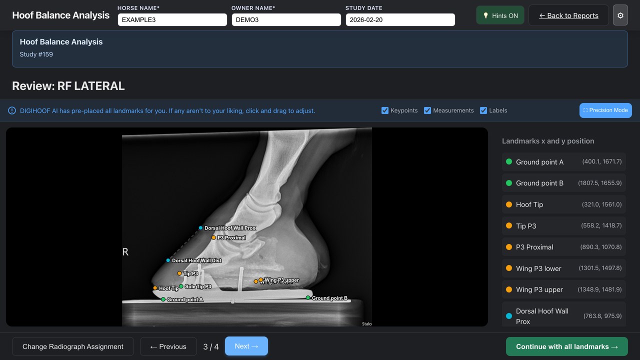
DigiHoof proposes the landmarks on your radiographs.
You review them, then share with the farrier, the client, and colleagues.
Five Regular Hoof Balance studies free on every new veterinarian account. No card required. Signup asks for your veterinary licence details.
What one study gives you
Open the study and the points are on the radiograph. Check each one and drag any that are off.
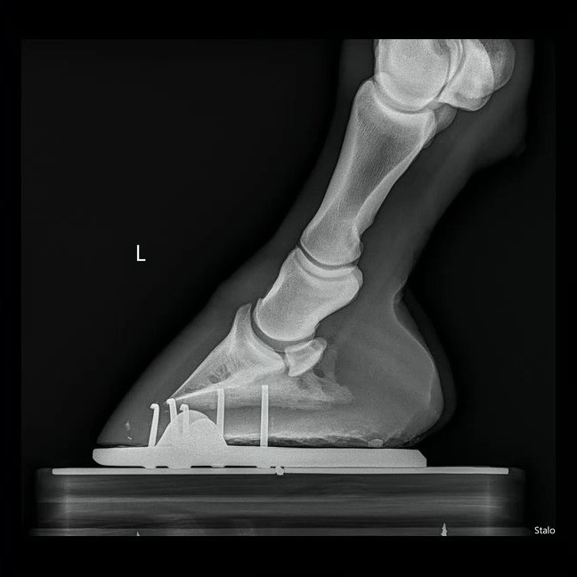
Lateral
DP
Scaled from the pixel spacing recorded in the DICOM. Move a landmark and the values are recalculated.
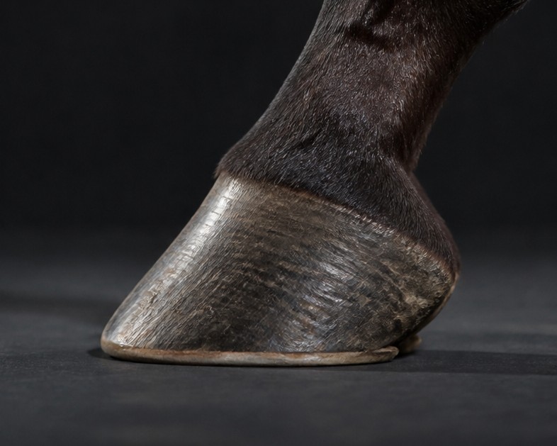
One page per foot: the marked radiographs beside your hoof photographs, the shoeing date, and your notes.
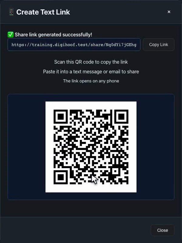
Text or email it to the farrier, the client, or a colleague. They do not need a DigiHoof account.
The structured measurements remain after the image files expire, while your account is active, so the next study can be set beside this one.

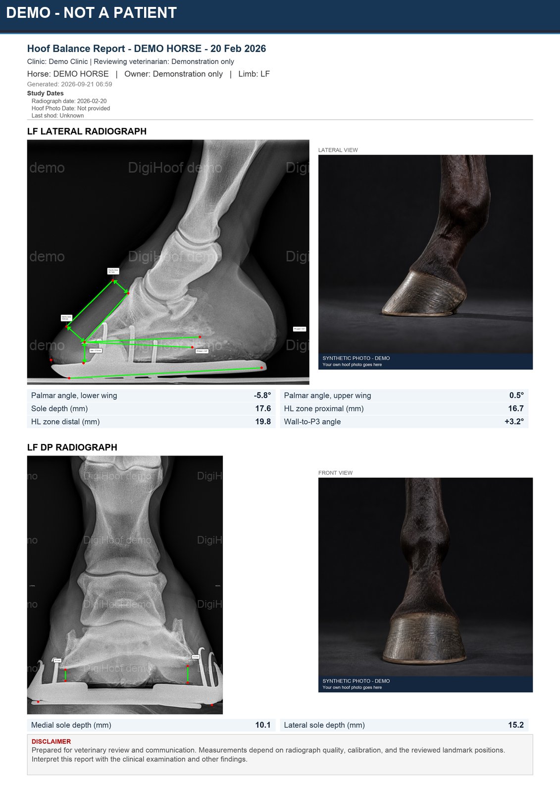
Real DigiHoof output from a demonstration study. Horse and owner names are placeholders. The hoof photographs are synthetic and labelled; your reports carry your own photographs.
What it does not do: DigiHoof does not assign a diagnosis, and it is not image storage. Source images and generated reports are temporary, and the study page shows their deletion dates. Download any files required for the permanent clinical record. Retention details →
For trimming and shoeing
5 free Regular Hoof Balance studies
For following lamellar zone measurements over time
Paid Membership required from the first LLZ study
Every new veterinarian account includes five free successfully analyzed Regular Hoof Balance studies. No card is required for those five. Membership is $15 USD per month per veterinarian and includes continued Regular studies plus LLZ.
The review screens
Lateral and DP views
Review palmar angle, sole depth, horn-lamellar zone, P3 rotation, and DP balance measurements.
See Hoof Balance measurements →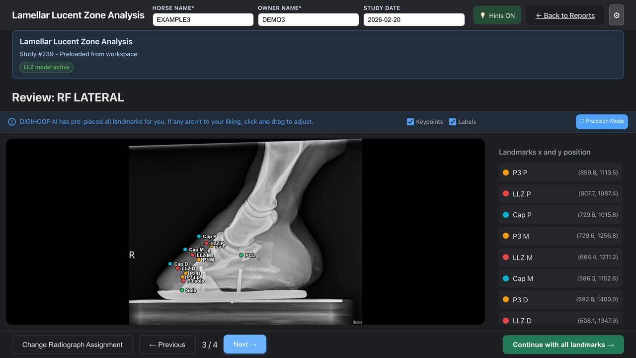
Lateral and DP views
Review proximal, middle, and distal LLZ widths, PCL, ratios, sole depth, and related hoof-wall measurements.
See LLZ measurements →DigiHoof proposes landmarks and calculates from the points you approve. It does not assign a diagnosis.
Built by Dr Alexander Daniel, BVetMed MS DACVS — a board-certified equine surgeon — and used in clinical practice. About DigiHoof →
The workflow
Drop DICOM files, a folder, or a ZIP. Hoof photos attach to the same study.
Check every proposed landmark. Move any point that does not match the radiograph. Measurements recalculate.
Save the PDF for the record. Send a link that opens on a phone.
Walkthroughs
Real screens. De-identified demonstration study. Captions included.
Hoof Balance · 1:56
LLZ · 2:25
Membership is $15 USD per month per veterinarian. It includes continued Regular Hoof Balance studies and LLZ. LLZ requires Membership from the first LLZ study.
Press Esc or click outside the image to close