Lamellar Lucent Zone analysis

Measure the Lamellar Lucent Zone (LLZ) on calibrated hoof radiographs.

DigiHoof proposes LLZ landmarks on lateromedial and dorsopalmar views. The veterinarian reviews and corrects the landmark geometry before accepting widths, ratios, or related measurements.

Primary LM outputs Proximal, middle, distal LLZ
Normalized output LLZmid:PCL ratio
Clinical control Editable, veterinarian-reviewed landmarks
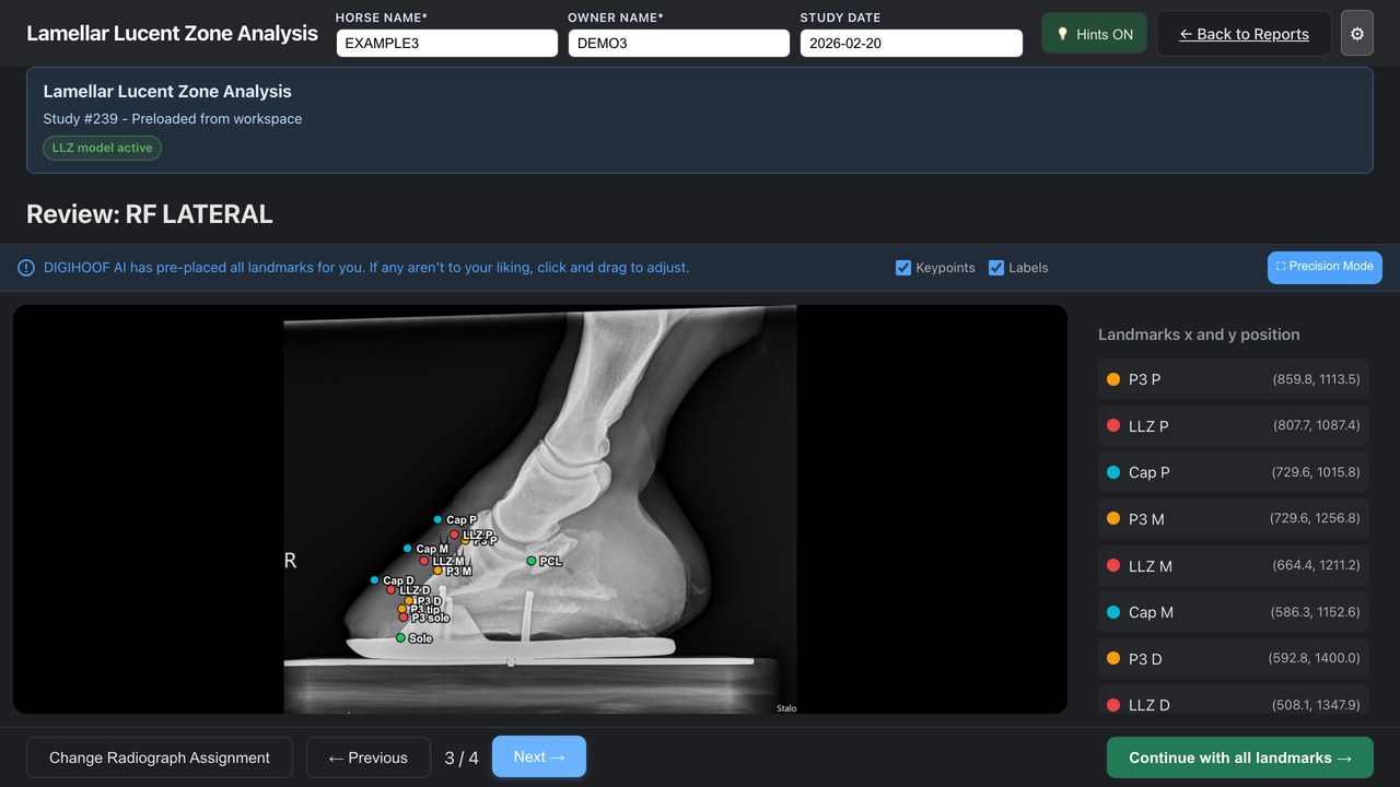
DigiHoof Lamellar Lucent Zone screen showing a de-identified lateromedial radiograph with editable LLZ landmarks
De-identified LLZ training study Genuine radiograph with synthetic horse and owner identities.

LLZ landmark control

Review the LLZ and reference landmarks before accepting a ratio.

The LLZ workspace displays the proposed LLZ, hoof-capsule, P3, sole, and PCL points on the source radiograph. Move an inaccurate point before DigiHoof recalculates the related widths and ratios.

Watch the LLZ walkthrough →

Calculated outputs

LLZ measurements are reported by level and projection.

Lateral LLZ widths are calculated perpendicular to the dorsal P3 reference axis using the final approved landmarks. Ratio outputs reduce, but do not eliminate, the effects of magnification and projection.

LM

Lateromedial projection

Widths, ratios, angles, and sole depth

  • LLZ widthProximal, middle, and distal levels
  • Palmar cortical length of P3PCL
  • LLZmid:PCLMiddle LLZ divided by PCL
  • LLZ:DHW-PdLevel-specific dorsal wall–P3 ratios
  • Inner and outer hoof wall–P3 anglesBest-fit dorsal reference lines
  • Sole depthP3 tip to solar surface
DP

Dorsopalmar projection

Medial and lateral measurements

  • Medial LLZ widthMedial dorsal wall–P3 region
  • Lateral LLZ widthLateral dorsal wall–P3 region
  • Medial and lateral sole depthSeparate solar measurements
  • Medial and lateral hoof wall widthReported by side
Side-to-side values require positioning review.

Rotation, obliquity, and marker placement can change the apparent medial/lateral relationship.

Image requirements

LLZ visibility and calibration must be adequate.

  • Correctly identified lateromedial or dorsopalmar projection
  • Appropriate positioning and beam alignment
  • Diagnostic exposure and post-acquisition windowing
  • Visible dorsal hoof wall and distal phalanx margins
  • Valid calibration information
  • Correct laterality and marker placement

Landmark review

The calculated value follows the approved point.

DigiHoof does not treat a proposed landmark as final. The veterinarian can move LLZ, P3, hoof-capsule, sole, and PCL points before recalculation. Values should be excluded when the underlying structures cannot be identified reliably.

Clinical evidence and limitations

LLZ measurements support assessment; they do not establish a diagnosis alone.

Skelton and colleagues evaluated LLZ measurements on equine hoof radiographs from acute, subacute, and control groups. The study reported that magnification and obliquity affected absolute LLZ measurements but did not alter the evaluated LLZ ratios. The authors also identified the absence of histologic confirmation as a study limitation.

DigiHoof displays measurements and literature-derived reference context for veterinarian review. Diagnosis and case classification still require clinical signs, history, examination, image technique, and professional interpretation.

Serial LLZ studies

Ratios improve comparability but do not replace technique control.

Compare the same foot and projection using consistent positioning, calibration, windowing, landmark definitions, and method versions. Review the radiographs alongside the numerical change.

Review serial comparison and reporting →

Optional paid workflow

Add LLZ analysis for $15 USD/month.

Hoof Balance remains available without a subscription. LLZ service begins immediately after checkout and can be canceled at any time.