
Hoof Balance

Foot balance on the lateral and DP, measured from the points you approve.

Drop your laterals and DPs into the browser. DigiHoof places the foot-balance landmarks, you correct any that are off, and the reviewed values go into a report you can send. The veterinarian inspects and corrects the landmarks before using the outputs.

Each new veterinarian account includes five free Regular Hoof Balance studies with no card required. Membership is required for study six.

Views Lateromedial and dorsopalmar
Measurement basis Calibrated, editable landmarks
Clinical control Veterinarian reviews every result
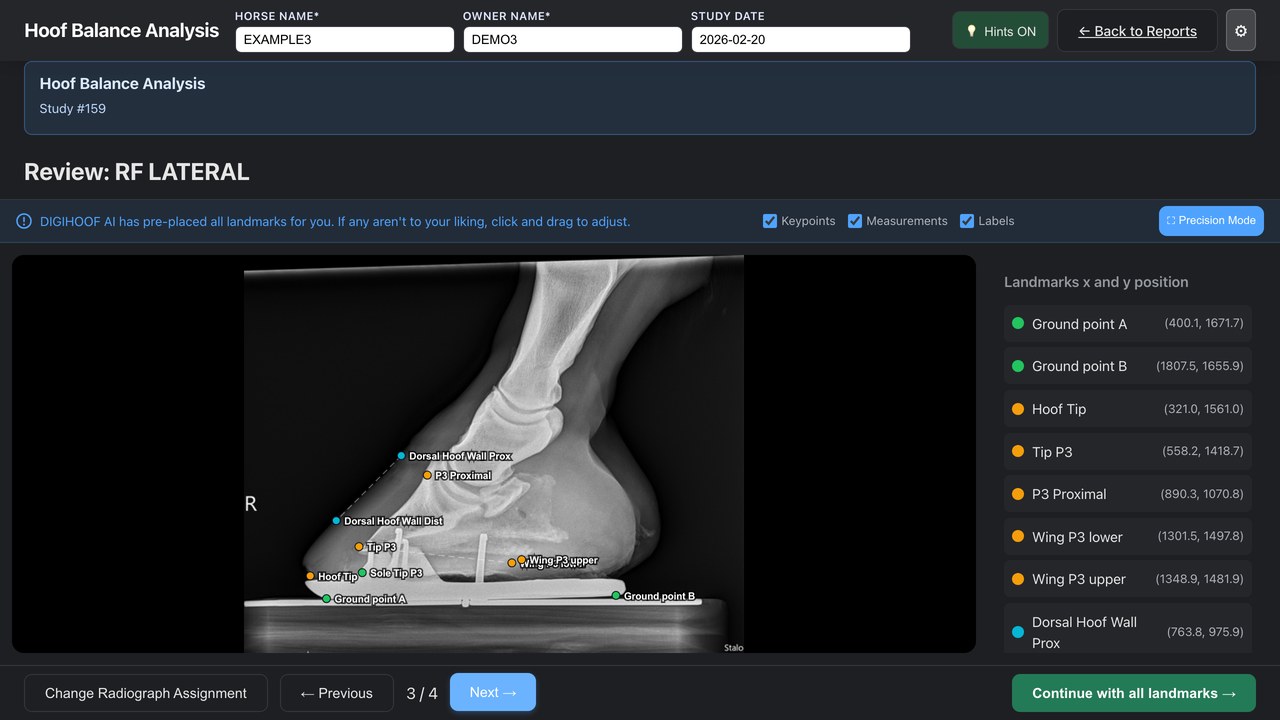
DigiHoof Hoof Balance screen showing a de-identified lateromedial radiograph with editable landmarks
De-identified Hoof Balance training study Genuine radiograph with synthetic horse and owner identities.

Landmark control

Every point is yours to move.

The review screen keeps the radiograph, named landmarks, and point coordinates together. Check the positioning, move any point that is not anatomically accurate, then continue to the calculated outputs.

Watch the Hoof Balance walkthrough →

Calculated outputs

What each view gives you.

Values are calculated from the landmark positions you approve and the pixel spacing recorded in the DICOM. Missing or unsuitable landmarks should not be reported.

LM

Lateromedial projection

Foot balance and sagittal measurements

  • Palmar angleUpper- and lower-wing references relative to ground
  • Sole depthDistal P3 tip to solar surface
  • Dorsal hoof wall–P3 distancesProximal and distal
  • Dorsal hoof wall–P3 angleRelationship of the two dorsal margins
  • P3 wing asymmetryVertical difference between wing references
  • Breakover relative to P3Ground reference relative to the P3 tip
  • Hoof tip–P3 tip distanceCalibrated linear distance
DP

Dorsopalmar projection

Medial and lateral solar measurements

  • Medial sole depthMedial P3 wing reference to solar surface
  • Lateral sole depthLateral P3 wing reference to solar surface
  • Side orientationConfirmed from the radiographic marker
Projection matters.

Rotation and beam alignment can alter apparent symmetry. DP measurements must be reviewed with positioning quality and marker placement in mind.

Before analysis

When not to trust the numbers.

  • Correct horse, limb, date, and projection
  • Appropriate positioning and beam alignment
  • Visible anatomical and ground-surface references
  • Valid calibration information
  • Correct medial/lateral marker placement on DP views

Before reporting

Check the image, then check the points.

  • Inspect the original radiograph at diagnostic quality
  • Correct proposed landmarks where required
  • Confirm the units and calculated values
  • Exclude outputs affected by image or calibration error
  • Interpret measurements with the complete clinical examination

Clinical limitation

DigiHoof cannot fix a poorly positioned radiograph.

DigiHoof standardizes landmark names, calculations, and reporting. It cannot make poorly positioned, oblique, uncalibrated, or nondiagnostic radiographs comparable. The veterinarian determines whether a measurement should be used.

Five free Regular studies per new veterinarian account.

No card is required. After the fifth successfully analyzed Regular study, DigiHoof Membership is $15 per month for continued Regular studies and LLZ.